Topic: Abdominal Pain
It’s a quiet Tuesday afternoon when you pick up the chart for Bed 6. The patient is a 42-year-old male, and the triage note is short: “Right flank pain. ?Kidney stone.”
You walk in expecting to see the classic kidney stone dance. You know the one, the patient writhing in agony, unable to find a comfortable position.
But that’s not what you see.
The patient is lying perfectly still. He looks flushed, miserable, and scared to move. When you ask him where it hurts, he grips his lower back and says, “It’s all right here in the flank. It started around my belly button about 12 hours ago, but now it’s all in my back.”
His vitals confirm he is sick. Temperature is 38.3°C and his heart rate is 102.

The Trap
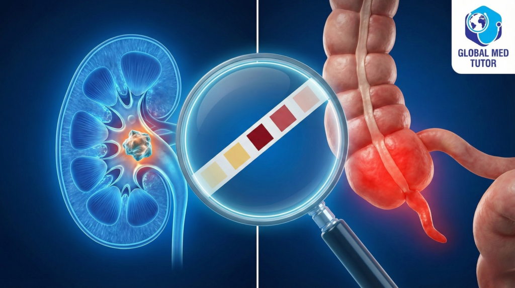
You order the standard workup. The bloods come back showing an infection with a white cell count of 14,000. Then the urine result arrives, and it seems to confirm exactly what the triage nurse thought.
- Red Blood Cells: Positive
- White Blood Cells: Positive
- Protein: Trace
It looks like a slam dunk. You have a patient with flank pain, fever, and blood in the urine. Your brain immediately jumps to Acute Pyelonephritis or an infected kidney stone. You are about to prescribe antibiotics and admit him under Urology.

Wait a Minute
Something bothers you about the history.
You ask him again, “You said the pain started around the belly button?”
“Yeah,” he nods. “Just a central stomach ache. Then it moved to the back.”
That sounds like the classic migration of appendicitis. But how can an appendix cause back pain and blood in the urine?
You decide to take a second look. When you press on his stomach, there is no guarding. It feels soft. But when you ask him to lift his right thigh against your hand—the Psoas Sign—he winces in pain.

The Anatomical Puzzle
This is where anatomy dictates the diagnosis.
This patient likely has a Retrocecal Appendix. This means his appendix is tucked behind the bowel, sitting right against the muscles of the back.
Because it is hidden in the back, it doesn’t irritate the front tummy muscles, so you don’t get that classic rock-hard abdomen. Instead, the inflamed appendix sits directly on the right ureter. It irritates the ureter from the outside, causing a little bit of blood and pus to leak into the urine.
Here is the subtle clue you need to watch for: Look closely at the urine numbers. In a true kidney stone, you often see massive amounts of blood. But here, the count is only 8 to 10 red cells per field. It’s abnormal, but not catastrophic. That is the sign of “sympathetic inflammation” from the nearby appendix, not a stone.

The Verdict
You bypass the renal ultrasound and order a CT scan. It reveals a swollen, fluid-filled appendix tucked deep behind the colon. There is no kidney stone.
The patient goes for a laparoscopic appendectomy and recovers perfectly. Urology never needed to be involved.
The Tutor’s Takeaway
1. Don’t be fooled by the dipstick It is a documented fact that up to 40% of appendicitis patients have abnormal urine findings. The inflammation near the ureter mimics a urinary tract infection.
2. Check the count Low-level hematuria usually points to the appendix. Massive hematuria points to a stone.
3. The Flank Pain Rule If the pain started centrally and moved to the back, think Retrocecal Appendicitis first. A kidney stone almost never starts at the belly button.
Need help mastering these subtle signs? Don’t rely on guesswork.
Download my Adult History Taking Script to ensure you catch that crucial migration of pain history every time, or
Book a 1-on-1 Free Clinical Reasoning Session with us to practice cases just like this.
References
- Paulson EK, Kalady MF, Pappas TN. Suspected Appendicitis. The New England Journal of Medicine. 2003.
- Avci MA, Akgün C, Uyanik MS. The Relationship Between Abnormal Urinalysis Findings and Appendicitis Location. International Journal of Colorectal Disease. 2023.
- Ong EM, Venkatesh SK. Ascending Retrocecal Appendicitis Presenting With Right Upper Abdominal Pain. World Journal of Gastroenterology. 2009.
- Kim S, Lim HK, Lee JY, et al. Ascending Retrocecal Appendicitis: Clinical and Computed Tomographic Findings. Journal of Computer Assisted Tomography. 2006.